Scientific overview
Idiopathic pulmonary fibrosis (IPF) and chronic obstructive pulmonary disease (COPD) disrupt lung homeostasis through different pathological processes. We reanalysed the published GSE136831 single-cell RNA-sequencing atlas to distinguish disease-associated shifts in recovered cell composition from gene-expression differences within individual populations. Across 307,650 cells and 78 clinical sample identifiers, IPF showed increased myofibroblast and reduced alveolar type II (ATII) representation. Pseudobulk analyses revealed substantial fibroblast and macrophage transcriptional differences, alongside heterogeneity within ATII cells. Pathway and marker analyses further identified an IPF-associated macrophage cell-cycle transcriptional phenotype, while exposing uncertainty about the cellular origin of some transcripts.
Establishing the analytical foundation
We retained the published Adams et al. annotations for 38 lung cell populations rather than claiming new cell-type discovery. The dataset comprised 95,303 Control cells (28 clinical samples), 144,404 IPF cells (32) and 67,943 COPD cells (18). Clinical sample identifiers were the units of biological inference, although their one-to-one correspondence with unique participants has not been independently verified; individual cells were not treated as independent replicates.
Figure 1. Clinical-sample distribution and recovered cell counts across disease groups.
We inspected transcript detection and mitochondrial RNA fractions to characterize variation in sample quality. These measures cannot rule out differential anatomical sampling, dissociation or capture efficiency.
Figure 2. Sequencing and cell-quality metrics across clinical samples.
The recovered lung cellular landscape
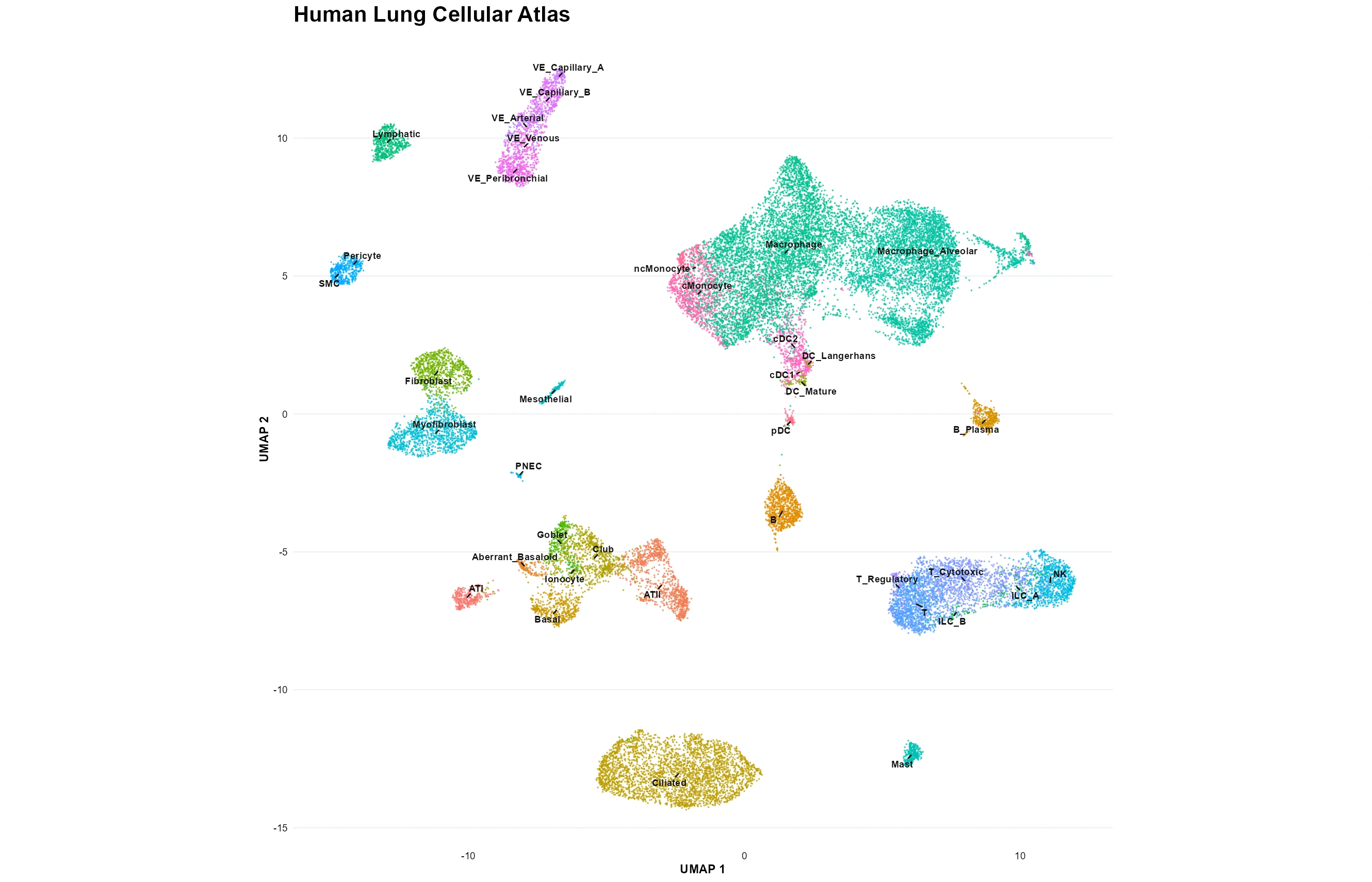
The published embedding placed 38 annotated epithelial, stromal, vascular and immune populations in a common transcriptional landscape. Disease-coloured UMAPs showed the distribution of recovered cells, not tissue abundance; lineage-marker expression provided supporting, but not independent experimental, evidence for the source annotations.
Figure 3. Source-annotated human lung cellular atlas.
Figure 4. Control, IPF and COPD cells across the shared lung embedding.
Figure 5. Canonical marker expression across annotated lung populations.
The atlas established which populations were observed. We next asked which changed consistently in representation across clinical samples.
Cellular composition identifies stromal and epithelial differences
We compared per-sample recovered fractions for 38 populations across IPF versus Control, COPD versus Control and IPF versus COPD (114 tests; Benjamini-Hochberg adjustment within comparison). In IPF, myofibroblasts increased from 0.26% to 2.02% (+1.76 percentage points; FDR = 0.00059), whereas separately annotated fibroblasts changed from 0.84% to 1.11% without statistical significance (FDR = 0.415). ATII representation declined from 2.11% to 0.30% (-1.81 percentage points; FDR = 0.0055). Aberrant basaloid, goblet and ciliated populations also differed significantly. These are relative fractions among recovered cells, not histological estimates of cell loss or expansion.
Figure 6. Clinical-sample variation in recovered lung cell fractions.
Two observations motivated focused investigation: the increase in myofibroblasts despite a nonsignificant change in fibroblast representation, and the lower recovery of ATII cells. Selection also reflected biological relevance and sample adequacy, not an exhaustive ranking of all 38 populations.
Fibroblasts reveal molecular remodeling beyond cell proportions
Fibroblasts contribute to extracellular-matrix organization, but their recovered fraction did not change significantly. Could their transcription nevertheless differ in IPF? Clinical-sample raw-count pseudobulk analysis using edgeR quasi-likelihood testing included 31 eligible samples and 6,045 genes. Of these, 405 met FDR < 0.05 and 162 additionally met |log₂ fold change| ≥ 1.
Figure 8. IPF-associated fibroblast differential expression.
To test dependence on individual samples, we repeated the analysis after excluding each eligible clinical sample in turn. All 20 prioritized genes retained their effect direction and FDR significance, although SULF1, SH3BGRL and TPM1 were less stable under the additional twofold-effect criterion.
Figure 9. Leave-one-clinical-sample-out stability of fibroblast associations.
Ranked-gene Hallmark enrichment showed negative oxidative-phosphorylation and MYC-target signals and positive coagulation and mitotic-spindle signals in IPF fibroblasts. These reflect coordinated RNA patterns, not direct measurements of metabolism, coagulation or proliferation.
Figure 10. Hallmark enrichment in IPF-associated fibroblast expression.
Thus, extensive transcriptional differences can coexist with a nonsignificant compositional change. The independently observed ATII reduction prompted a complementary epithelial investigation.
ATII cells exhibit reduced recovery and transcriptional heterogeneity
ATII cells support surfactant production and alveolar maintenance. Their lower recovered representation in IPF motivated within-population pseudobulk testing, which identified 90 genes at FDR < 0.05, including 73 with |log₂ fold change| ≥ 1. This does not establish impaired surfactant secretion or failed tissue repair.
Figure 11. IPF-associated differential expression in broad ATII cells.
The source annotations further distinguish high-surfactant and low-surfactant ATII groups by relative expression of surfactant-associated genes. These are transcriptomic categories, not proof of functional health or injury. Because broad ATII differences might reflect changing subgroup representation, within-group expression changes or both, we examined subgroup recovery before comparing expression-effect estimates.
Figure 13. High- and low-surfactant ATII representation across clinical samples.
Figure 14. IPF-associated expression estimates in broad and surfactant-defined ATII groups.
Sparse ATII recovery limited inference: Control/IPF eligibility fell from 16/13 samples with at least 10 cells to 15/9 at 15 cells and 13/7 at 20 cells. A historical 17-gene recurrent set has not yet been confirmed against the joint FDR < 0.05 and |log₂ fold change| ≥ 1 criteria in all three complete fits. The defensible result is IPF-associated ATII transcriptional heterogeneity with sensitivity to sample-eligibility thresholds.
Having examined stromal and epithelial compartments, we returned to the original composition evidence to investigate immune-cell differences, particularly whether macrophage transcription distinguishes IPF from COPD.
Macrophage transcription distinguishes disease-associated states
Macrophages regulate pulmonary immune surveillance and responses to injury. Their recovery differed across diseases, particularly in comparisons involving COPD. Broad alveolar-macrophage pseudobulk profiles included 26 Control, 31 IPF and 17 COPD samples. IPF versus Control yielded 6,902 FDR-significant genes (643 with |log₂ fold change| ≥ 1); COPD versus Control yielded 1,073 (259); and direct IPF versus COPD yielded 5,871 (588).
Figure 15. Broad alveolar-macrophage differential expression across disease contrasts.
A difference in significance against Control does not itself establish a difference between diseases. We therefore examined the direct IPF-versus-COPD test alongside the corresponding IPF-versus-Control and COPD-versus-Control effect estimates.
Figure 16. Comparison of IPF-versus-Control and COPD-versus-Control macrophage expression effects, interpreted with direct IPF-versus-COPD testing.
Could these extensive results reflect changes in the mixture of macrophage subgroups? The published annotations distinguish core, cell-cycle-associated and atypical macrophage transcriptomic groups. We inspected their recovered fractions, then repeated IPF-versus-Control testing within core macrophages.
Figure 17. Source-defined macrophage transcriptomic groups across disease cohorts.
Core macrophages retained 6,531 FDR-significant genes, including 490 meeting the twofold-effect criterion. Broad and core effect estimates were strongly concordant, indicating that the expression pattern cannot be explained solely by the separately annotated subgroup mixture; residual heterogeneity and confounding remain possible.
Figure 18. IPF-associated expression differences in core macrophages.
Figure 19. Concordance of broad- and core-macrophage disease-effect estimates.
Pathway enrichment motivates a targeted cell-cycle test
Hallmark enrichment of ranked macrophage results identified positive E2F-target, MYC-target, G2M-checkpoint and oxidative-phosphorylation signatures in IPF. These patterns remained broadly similar after adjustment for recovered macrophage count, supporting robustness to this measured sampling characteristic, not proving metabolic activity or cell division.
Figure 20. Core-macrophage pathway enrichment with and without cell-count adjustment.
The cell-cycle transcriptional signal motivated an independent marker-level question: do more macrophage-labelled cells jointly detect MKI67 and TOP2A? Co-detection occurred in 349/23,905 Control, 1,324/27,403 IPF and 207/23,250 COPD cells. Clinical-sample-level quasibinomial modelling estimated an IPF-versus-Control odds ratio of 3.43 (95% CI 2.39-4.92; FDR = 2.97 × 10⁻⁸), with consistent direction across 60 leave-one-sample-out refits.
Figure 21. MKI67/TOP2A co-detection in macrophage-labelled cells.
This strengthens evidence for a cell-cycle-associated RNA phenotype, not experimentally verified macrophage proliferation. The breadth of macrophage transcriptional differences also required scrutiny of unexpected lineage-associated signals.
Unexpected transcripts delimit biological interpretation
Macrophage-labelled cells detected platelet-associated PPBP/PF4 and epithelial-associated MMP7/KRT19 RNA. Median core-macrophage PPBP detection was 0.17% in Control, 15.30% in IPF and 0.54% in COPD. Possible sources include intrinsic expression, phagocytosed material, ambient RNA and multiplets; detection alone cannot distinguish them or demonstrate platelet-protein synthesis by macrophages.
Figure 22. Platelet- and epithelial-associated transcript detection in macrophage-labelled cells.
These findings do not negate the macrophage analysis, but they limit cell-intrinsic interpretation and illustrate why conspicuous transcriptomic associations require source-of-RNA scrutiny.
Conclusion
IPF and COPD exhibit distinct cellular and transcriptional disease associations rather than a uniform shift in the lung atlas. In IPF, increased recovered myofibroblast and reduced ATII representation accompany fibroblast and epithelial transcriptional differences; macrophages show extensive disease-associated expression divergence, including a robust cell-cycle-associated RNA phenotype. The persistence of broad macrophage signals in core cells strengthens their interpretation, whereas unexpected platelet- and epithelial-associated transcripts constrain claims about cellular origin. Together, the evidence resolves stromal, epithelial and immune patterns at clinical-sample level without conflating recovered composition, gene expression and demonstrated mechanism.